When you ask what does a hair follicle look like, the answer involves a complex structure hidden beneath the skin that determines the health, texture, and growth of every strand of hair. Knowing the visual characteristics of a hair follicle helps clinicians diagnose scalp conditions, guides patients in choosing appropriate hair‑restoration treatments, and demystifies common myths about hair loss. In fact, studies show that up to 70 % of people experience some form of follicular concern during their lifetime, making awareness essential.
This article breaks down the anatomy of the follicle, explores how it appears under different magnifications, examines the factors that alter its look, and highlights when changes signal a medical issue. Whether you are considering a hair transplant at Gold City Medical Center or simply curious about your scalp health, understanding follicle appearance empowers you to make informed decisions.
We will walk through the layers from the hair bulb to the surrounding dermal sheath, illustrate typical microscopic images, and discuss practical assessment methods used by professionals. By the end, you will have a clear mental picture of the structure that fuels every hair on your head.
Hair Follicle Anatomy: Core Structures Explained
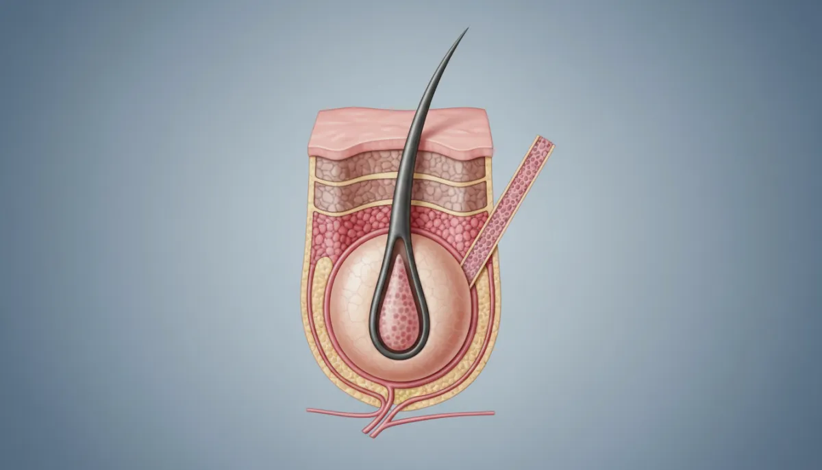
The hair follicle is a dynamic mini‑organ composed of several distinct parts, each contributing to the overall appearance and function. Below is a concise overview of the main components:
- Hair Bulb: The rounded base where cells divide rapidly during the anagen phase.
- Hair Papilla: A cluster of capillaries that supplies nutrients to the bulb.
- Hair Root: The portion of the hair shaft embedded within the follicle.
- Inner Root Sheath (IRS): Guides the emerging hair shaft and protects it.
- Outer Root Sheath (ORS): Connects the follicle to the surrounding epidermis.
- Arrector Pili Muscle: Tiny muscle that contracts to raise the hair (goosebumps).
These elements are arranged in concentric layers, creating a tubular shape that can be visualized in cross‑section. The following table compares the key features of each part:
| Component | Location | Primary Function | Visual Cue |
|---|---|---|---|
| Hair Bulb | Base of follicle | Cell proliferation for hair growth | Rounded, pigmented |
| Hair Papilla | Within bulb | Blood supply | Bright, vascular |
| Inner Root Sheath | Surrounds hair shaft | Guides shaft | Layered, translucent |
| Outer Root Sheath | Extends to epidermis | Structural support | Thick, continuous |
| Arrector Pili Muscle | Attached to ORS | Hair erection | Fine, fibrous |
Understanding these layers clarifies why a healthy follicle appears as a well‑organized, multi‑layered unit when examined under magnification. Any disruption—such as inflammation or scarring—will alter this orderly look, often visible as irregularities in the sheath or bulb shape.
How Hair Follicles Appear Under the Microscope
When clinicians answer the question what does a hair follicle look like under a microscope, they typically use either a light microscope or a scanning electron microscope (SEM). Each provides a distinct visual perspective:
- Light Microscopy: Offers a 2‑D view of stained sections, highlighting cellular detail.
- Scanning Electron Microscopy: Delivers a 3‑D surface view, revealing the follicle’s external contour.
- Confocal Microscopy: Enables depth‑stacked images for precise layer differentiation.
In a standard light‑microscope slide, the hair bulb appears as a dark, densely packed region, while the papilla shows up as a lighter, vascular core. The inner and outer root sheaths are distinguishable by their staining intensity, and the arrector pili muscle appears as a thin, fibrous band attached to the outer sheath.
Below is a simple comparison of the visual characteristics you can expect from each technique:
| Technique | Resolution | Typical Appearance | Best Use |
|---|---|---|---|
| Light Microscopy | 0.2 µm | Cross‑sectional layers, stained contrast | Routine diagnostic biopsies |
| Scanning Electron Microscopy | 5 nm | 3‑D surface topology, fine texture | Research on follicle surface changes |
| Confocal Microscopy | 0.1 µm | Layered depth images, fluorescent labeling | Live‑cell studies, dynamic processes |
These imaging modalities help answer the primary question by providing visual evidence of normal versus abnormal follicle morphology. For example, a healthy follicle shows a smooth, rounded bulb, whereas a follicle affected by inflammation may appear swollen or irregular.
Factors Influencing Follicle Appearance: Health, Age, and Genetics
Many variables can alter the visual characteristics of a hair follicle, making the answer to what does a hair follicle look like highly individual. The most influential factors include overall health, hormonal balance, age‑related changes, and genetic predisposition.
Health Conditions such as thyroid disorders, anemia, or chronic inflammation can cause the follicle to appear smaller or less vascular. Nutrient deficiencies—especially in iron, zinc, and biotin—may lead to a thinner hair shaft and a less pronounced bulb.
Hormonal Influences play a crucial role. Elevated dihydrotestosterone (DHT) can shrink follicles, a process known as miniaturization, which is visually evident as a reduced bulb diameter and shortened hair shaft.
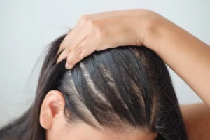
Veroudering naturally reduces the number of active follicles. Older follicles often display a flattened bulb, reduced papilla size, and a thinner outer root sheath. These changes contribute to the gradual thinning seen in many adults.
Genetics determines baseline follicle density and the propensity for conditions like androgenic alopecia. People with a family history of hair loss may notice early signs of follicular miniaturization, visible under magnification as a shift from thick, pigmented bulbs to finer, less pigmented structures.
The table below summarizes how each factor typically manifests in follicle appearance:
| Factor | Visual Change | Clinical Implication |
|---|---|---|
| Nutrient Deficiency | Smaller bulb, pale papilla | Potential hair thinning, slower growth |
| Elevated DHT | Miniaturized bulb, shortened shaft | Androgenic alopecia progression |
| Veroudering | Flattened bulb, thinner sheath | Reduced density, increased shedding |
| Genetische aanleg | Early miniaturization, variable density | Higher risk of patterned hair loss |
By recognizing these visual cues, clinicians can tailor treatment plans—ranging from nutritional supplementation to targeted hair‑restoration procedures—ensuring each patient receives care that matches the underlying follicular condition.
Common Concerns: When Follicle Appearance Indicates a Problem
Understanding what does a hair follicle look like in a healthy state helps you spot warning signs early. The most common visual abnormalities include:
- Ontsteking: Swollen sheath, reddened papilla, and irregular bulb shape.
- Scarring (Cicatricial Alopecia): Fibrotic tissue replaces normal follicular structures, leading to a flattened, scar‑like appearance.
- Folliculitis: Presence of pus or bacterial colonies visible as bright spots within the follicle.
- Miniaturization: Progressive reduction in bulb diameter and hair shaft thickness.
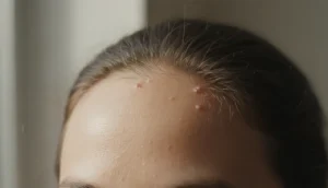
- Keratin Plugging: Accumulation of keratin debris that blocks the follicular opening, seen as a dark plug at the surface.
These conditions often manifest clinically as itching, redness, or noticeable hair loss. A dermatologist may perform a scalp biopsy to directly observe the follicle’s morphology, confirming whether the visual changes correspond to a specific pathology.
Early detection is key. For instance, identifying inflammation before it progresses to scarring can preserve follicular integrity and improve the success rate of subsequent hair‑restoration procedures.
Hair Follicle Assessment in Clinical Practice
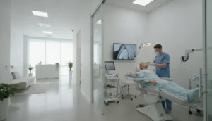
At Gold City Medical Center, evaluating the question “what does a hair follicle look like” is a systematic process that combines visual inspection, dermatoscopy, and, when needed, histological analysis. The workflow typically follows these steps:
- Visual Examination: Trained clinicians assess scalp texture, follicular density, and any visible lesions.
- Dermatoscopic Imaging: A handheld device magnifies the follicle surface, revealing details such as perifollicular scaling or vascular patterns.
- Biopsy & Histology: Small tissue samples are stained and examined under a light microscope to evaluate bulb size, papilla health, and sheath integrity.
- Laboratory Tests: Blood work may be ordered to rule out systemic causes affecting follicle appearance.
- Personalized Treatment Planning: Based on findings, options range from topical therapies and nutritional support to advanced hair‑transplant techniques.
Below is a concise checklist that clinicians use during a follicular assessment:
| Assessment Step | Tool Used | Key Visual Indicator |
|---|---|---|
| Visual Examination | Magnifying glass | Follicle density, surface lesions |
| Dermatoscopy | Dermatoscope | Perifollicular erythema, scaling |
| Histology | Light microscope | Bulb size, papilla vascularity |
| Blood Tests | Laboratory analysis | Iron, thyroid, hormone levels |
By following this structured approach, practitioners can accurately answer the patient’s curiosity about follicle appearance while delivering targeted, evidence‑based care.
Why Choose Gold City Medical Center
Gold City Medical Center combines cutting‑edge technology with a holistic approach to hair health. Our team of experienced dermatologists and hair‑restoration specialists uses advanced imaging and personalized treatment plans to ensure each follicle receives the care it needs. Whether you seek a detailed scalp analysis or a state‑of‑the‑art hair transplant, we deliver results grounded in scientific expertise and patient‑centered compassion.
Ready to see your follicles at their best? Contact Gold City Medical Center today for a comprehensive scalp evaluation and discover the path to healthier, fuller hair.
FAQ
What does a hair follicle look like under a microscope?
Under light microscopy the hair bulb appears dark and rounded, the papilla is lighter and vascular, while the sheaths show distinct staining.
What are the main parts of a hair follicle?
The primary components are the hair bulb, hair papilla, hair root, inner root sheath, outer root sheath, and the arrector pili muscle.
How does DHT affect hair follicle appearance?
Elevated DHT causes follicle miniaturization, seen as a reduced bulb diameter and shorter, thinner hair shaft.
What visual signs indicate an unhealthy hair follicle?
Inflammation, scarring, folliculitis, miniaturization, and keratin plugging are common visual abnormalities.
How can clinicians assess hair follicle health in practice?
Assessment combines visual inspection, dermatoscopy, and histological analysis when needed.