Many people wonder what does eczema look like, and recognizing its signs is the first step toward effective care. Eczema, also known as atopic dermatitis, presents with a wide range of visual cues that can vary by age, body part, and severity. By learning to spot these patterns early, you can seek appropriate eczema treatment and reduce discomfort.
In this article we will explore the typical appearance of eczema, how it differs on various parts of the body, and which forms—such as nummular eczema or weeping eczema—require prompt medical attention. Whether you are a parent noticing a rash on a newborn or an adult dealing with chronic flare‑ups, understanding the visual language of eczema empowers you to manage the condition confidently.
Recent studies show that up to 20 % of children experience eczema symptoms before the age of five, highlighting the importance of early detection. Below, we break down the most common visual presentations, explain why they occur, and offer practical guidance on when to consult a specialist at Gold City Medical Center.
Common Visual Patterns of Eczema
Eczema typically begins as dry, red patches that may become itchy, scaly, or cracked. The classic eczema rash often appears on the flexural surfaces—such as the inner elbows and behind the knees—but can manifest anywhere on the skin. Key characteristics include:
- Red or pink base color that may darken over time.
- Fine scaling or rough texture, especially after scratching.
- Small fluid‑filled blisters in acute flare‑ups.
- Thickened, leathery skin (lichenification) with chronic scratching.
Below is a comparison table that highlights how the rash evolves from acute to chronic stages:
| Stage | Appearance | Typical Symptoms |
|---|---|---|
| Acute | Red, inflamed patches with vesicles | Intense itching, burning |
| Sub‑acute | Dry, scaly lesions; some crusting | Persistent itch, mild soreness |
| Chronic | Thickened, hyperpigmented plaques | Reduced itching but stiff skin |
Recognizing these stages helps you gauge the severity and decide whether over‑the‑counter moisturizers are enough or a professional eczema treatment plan is warranted.
Eczema on Different Body Areas
The visual presentation of eczema can differ markedly depending on the location. Understanding these variations is essential for accurate self‑assessment.
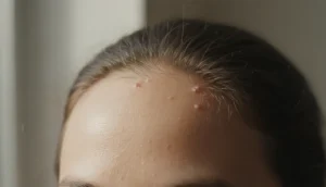
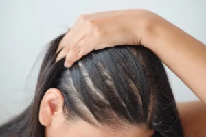
Face and Scalp
Facial eczema often appears as pale, dry patches on the cheeks, around the eyes, or on the forehead. The scalp variant may cause flaky, itchy dandruff‑like scales that can be mistaken for seborrheic dermatitis.
Hands and Feet
Hand eczema, sometimes called hand dermatitis, shows as red, cracked skin on the palms and fingers, often worsening with exposure to irritants. Foot eczema may involve the soles and can be confused with athlete’s foot, but it usually lacks the distinct fungal odor.
Infants and Babies
In newborns, eczema often presents as small, raised bumps (often called “baby eczema”) that may ooze clear fluid. These lesions frequently appear on the cheeks, scalp, and the creases of the elbows and knees.
Below is a quick reference list for common body‑specific signs:
- Face: Pale, dry patches, sometimes with crusting.
- Scalp: Flaky, itchy scales resembling dandruff.
- Hands: Cracked, painful skin on palms and fingers.
- Feet: Red, itchy soles, often mistaken for fungal infection.
- Infants: Small, weeping bumps on cheeks and scalp.
Identifying the exact location helps clinicians choose targeted therapies, such as gentle steroid creams for the face or barrier‑repair ointments for the hands.
Variants of Eczema and Their Distinct Appearances
Beyond the common atopic form, several eczema subtypes have unique visual signatures. Recognizing these can guide more precise treatment.
Nummular Eczema
Also called “discoid eczema,” nummular eczema presents as round, coin‑shaped patches that are often oozing and itchy. The lesions are typically well‑defined, with a yellowish crust in the center.
Dyshidrotic (Pompholyx) Eczema
This variant appears on the sides of the fingers and toes as tiny, fluid‑filled blisters that may burst and leave raw, painful skin.
Contact Eczema
Resulting from an allergic reaction to a substance, contact eczema shows up as localized redness, swelling, and sometimes blistering directly where the irritant touched the skin.
Comparison of key features:
| Variant | Typical Shape | Common Locations | Notable Features |
|---|---|---|---|
| Nummular | Round, coin‑shaped | Extremities, trunk | Oozing, crusted center |
| Dyshidrotic | Small vesicles | Sides of fingers & toes | Intense itching, blister burst |
| Contact | Irregular | Areas of exposure | Rapid onset after contact |
Understanding which variant you are dealing with can influence the choice of topical steroids, moisturizers, or avoidance strategies.
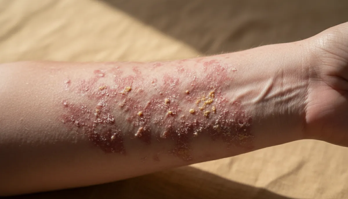
When Eczema Looks Infected or Weeping
Sometimes eczema becomes secondarily infected, changing its appearance dramatically. Recognizing these signs is crucial because infection requires prompt medical intervention.
- Yellow or greenish crusts over the rash.
- Increased warmth and swelling around the lesion.
- Pus‑filled blisters that may burst, leading to weeping skin.
- Fever or general feeling of illness.
This condition is often referred to as weeping eczema or infected eczema. If you notice any of these signs, it is advisable to see a dermatologist for possible oral antibiotics in addition to your regular eczema regimen.
Below is a quick checklist for identifying infection:
- Redness spreading beyond original borders.
- Exudate that is thick, yellow, or foul‑smelling.
- Rapid increase in itching or pain.
- Systemic symptoms such as fever.
Early treatment can prevent complications like cellulitis and reduce the risk of scarring.
How to Distinguish Eczema from Similar Skin Conditions
Several skin disorders can mimic eczema, making self‑diagnosis challenging. Knowing the distinguishing features helps you decide whether to manage at home or seek professional care.
Psoriasis vs. Eczema
Psoriasis often presents as well‑defined, silvery‑scale plaques, typically on the elbows, knees, and scalp. Unlike eczema, psoriasis plaques are usually less itchy and may bleed when scratched.
Contact Dermatitis vs. Atopic Dermatitis
Contact dermatitis is directly linked to exposure to an irritant or allergen and appears only where the skin contacted the substance. Atopic dermatitis tends to be more widespread and chronic.
Fungal Infections vs. Eczema
Fungal infections (tinea) often have a raised, ring‑shaped border with central clearing, and they may emit a mild odor. Eczema lesions are generally more uniform in color and lack the distinct ring.
Use this comparison chart to quickly assess key differences:
| Condition | Typical Shape | Scale Color | Itch Intensity | Common Triggers |
|---|---|---|---|---|
| Eczema | Irregular patches | Red, sometimes white | High | Genetics, allergens, stress |
| Psoriasis | Well‑defined plaques | Silvery | Moderate | Immune factors |
| Contact Dermatitis | Localized to contact area | Red, sometimes vesicular | Variable | Irritants, allergens |
| Fungal Infection | Ring‑shaped | White/cream | Moderate | Warm, moist environments |
If you are uncertain, a dermatologist at Gold City Medical Center can perform a skin examination and, if needed, a simple patch test or skin scraping to confirm the diagnosis.
Why Choose Gold City Medical Center
At Gold City Medical Center we combine integrative medicine with cutting‑edge dermatology to provide personalized care for eczema and related skin concerns. Our team of experienced specialists offers comprehensive assessments, tailored treatment plans, and ongoing support to help you achieve lasting relief. Whether you need prescription‑strength moisturizers, phototherapy, or advanced biologic therapies, we prioritize your comfort and skin health in a welcoming environment.
Ready to take control of your skin? Contact Gold City Medical Center today to schedule a personalized consultation and start your journey toward healthier, calmer skin.
FAQ
What does eczema look like in its early stages?
Early eczema appears as red, inflamed patches that may have small fluid‑filled blisters and intense itching.
How can I tell if my eczema is infected or weeping?
Infected or weeping eczema shows yellow/green crusts, increased warmth, swelling, pus‑filled blisters, and sometimes fever.
What are the visual differences between eczema and psoriasis?
Psoriasis shows well‑defined silvery plaques, while eczema has irregular red patches with intense itching.
Which type of eczema is common on the hands and feet?
Hand eczema (hand dermatitis) and foot eczema commonly affect the palms, fingers, and soles.
How does nummular eczema appear on the skin?
Nummular eczema shows round, coin‑shaped patches that are often oozing and have a yellowish crust in the center.
When should I seek professional eczema treatment for my child?
If the rash is widespread, persistent, painful, shows signs of infection, or interferes with sleep, see a dermatologist promptly.